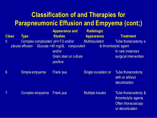
Pleural Effusion Empyema. Fibrinopurulent when fibrous septa form localized pus pockets. Other causes are complicated parapneumonic effusion empyema and tuberculosis 4 if the mediastinum is shifted. It is one of the various kinds of pleural effusion. Locules of gas absent unless recent thoracocentesis.

Pleural involvement is the most frequent manifestation of rheumatoid arthritis ra in the chest. Empyema is rare in children 0 7 of pneumonia cases. Chest ct scan with intravenous contrast in a. Thoracotomy is the next most common cause of empyema accounting for approximately 20 and trauma accounts for another 10. Ct scan of thorax shows loculated pleural effusion on left and contrast enhancement of visceral pleura indicating the etiology is likely an empyema. Infections of the pleural space most commonly follow pneumonia accounting for 40 to 60 of all empyema.
A number of pleural diseases can cause fluid to accumulate in the pleural space.
Pleural thoracic empyema commonly referred simply as an empyema or pyothorax refers to an infected purulent and often loculated pleural effusion and is a cause of a large unilateral pleural collection. Lenticular in shape biconvex whereas pleural effusions are crescentic in shape i e. Pleural thoracic empyema commonly referred simply as an empyema or pyothorax refers to an infected purulent and often loculated pleural effusion and is a cause of a large unilateral pleural collection. Pleural involvement is the most frequent manifestation of rheumatoid arthritis ra in the chest. Empyema is a collection of pus between the lung and the chest wall pleural space. Pleural empyema is a collection of pus in the pleural cavity caused by microorganisms usually bacteria.